

 RTI Act
Early Detection of Oral Cancer
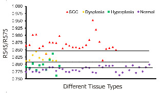
Fig. 1. Combined DR spectral ratio R545/R575 scatter plot discriminating different grades of cancer from 48 sites in 29 patients compared with the mean normal values in 36 healthy volunteers.
Oral cavity cancer represents a significant health problem owing to its high rate of incidence in India as well as the world over. Nearly 85% of the oral cancers are categorized as squamous cell carcinoma. As with all cancers, the prospect of curability is better when the malignancy is detected in an early stage. Therefore, regular screening of apparently healthy people is rewarding as it permits diagnosis before visible symptoms develop. Premalignant dysplastic lesions are routinely detected through invasive biopsy followed by histopathological examination, which is considered as the gold standard. However, histopathological examination has several drawbacks. A suspicious lesion that is premalignant at some part may not be malignant at another location. Therefore, biopsy from one location of the lesion may not be a representative of the entire lesion. Also, the resemblances of tissue inflammation and irritation with premalignant oral mucosal alterations and field cancerous changes are often challenging. This usually leads to random or repeated biopsies causing discomfort to the patients. Multiple stage sample preparations and processes are also time consuming and increase pathological costs.
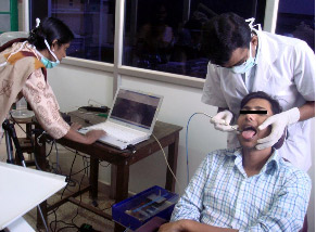
Fig. 2. A clinical trial to detect oral pre-cancer using the LIFRS device developed in the lab.
The prevalence of various optical spectroscopy techniques has been increasing and is getting greater recognition and acceptance these days owing to their noninvasive nature of tissue characterization. Among these techniques the potential of laser-induced tissue autofluorescence and diffuse reflectance (DR) are immense, but not yet fully explored for detection of precancerous lesions (Fig. 1).
The Biophotonics laboratory of the CESS has developed a LIFRS system for point monitoring and diagnosis of oral cavity cancer from autofluorescence and DR spectral features. A multi-spectral DR R545/R575 ratio imaging system based on EMCCD camera was used in a clinical trial to study the tissue morphology across the entire lesion in real time for identifying the most malignant site in a lesion for biopsy and in oral cancer detection (Fig. 2).