

 RTI Act
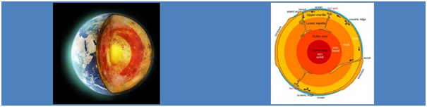
Electron Probe Micro Analyser (EPMA) Facility
NCESS has installed a Fifth Generation EPMA (Electron Probe Micro Analyser) SXFive-Tactis from CAMECA, France. EPMA is an instrument primarily used for the in situ non-destructive chemical analysis of minute solid samples. The ability of EPMA to acquire precise, quantitative elemental analyses at very small "spot" sizes (1-2 microns), mainly by wavelength-dispersive spectroscopy (WDS) combined with the ability to create detailed images of the sample, makes it possible to analyze geological materials in situ and to resolve complex chemical variation within single phases. NCESS-EPMA will be primarily used for (i) chemical analysis of geological materials, (ii) analysis of individual phases (e.g., igneous and metamorphic minerals), (iii) U-Th-Pb geochronology of minerals such as monazite, zircon etc.
Instrumentation
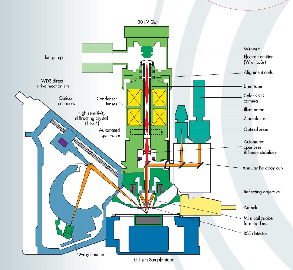
NCESS-CAMECA SXFive-Tactis is equipped with five wave length dispersive spectrometers, BSE detectors, SE detectors, cathodoluminescence and sophisticated visible light optics. The self-biased electron gun of the instrument has the provision for both W and LaB6 filaments. The instrument is configured in such a way so that operation as well as basic imaging and data processing are made easy for having intuitive touch screen interface. In expert mode, the interface is designed to have benefit from a full complement of different tool parameters and software options. The system involves a high degree of automation with the capacity of prolonged unattended analytical schedules with high analytical precision, accuracy and reproducibility with minimum down-time, high quality imaging at high spatial resolution including multi-element x-ray mapping. Vertical WDSs are compatible to have suitable combinations in high sensitivity diffracting crystals (e.g. LTAP, LPC1, LLIF and LPET) as well as six diffracting crystals such as TAP, LIF, PET, PC0, PC3 and PC2. Another advantage of this instrument is that it is equipped with a fully integrated Electron Dispersive Spectrometer hypermapping module making data processing and analysis faster and easier. SX- Five with LaB6 Column: Accelerating Voltage: 15KV and 20 KV; Beam Current: 20 uA onwards; Beam Stability: +-0.5% per hour @ 20kV, 20nA.
Capabilities
Qualitative analysis: WDS Spectrum-to find out the elements present. Wavelength Dispersive Spectrometry is acknowledged as the method of choice for high precision quantitative microanalysis. The SXFive-Tactis spectrometers have the highest resolution and, as a result high peak to background ratios, thereby providing high sensitivities. The absolute spectral positioning is provided by optical encoders attached to the system. Only one peak measurement is needed to calibrate the entire spectrometer range.
Quantitative analysis: Accurate quantification of chemical composition of any solid material. SXFive-Tactis delivers the true quantitative analysis. The quantitative data can be displayed such as wt%, oxide wt%, cations. See the table below for our precision:
|
Cameca Silicate Std (Alamandine) |
sample |
Cameca Sulphide Std (Pyrite) |
sample |
Uranium Oxide Std |
sample |
|||
|
SiO2 |
37.87 |
37.51 |
S |
53.06 |
52.86 |
Al2O3 |
0.29 |
0.07 |
|
TiO2 |
0.07 |
0.04 |
Fe |
46.51 |
45.89 |
UO2 |
89 |
89.28 |
|
Al2O3 |
21.36 |
21.16 |
Co |
0.5 |
0.35 |
SiO2 |
2.3 |
7.23 |
|
Cr2O3 |
0.0 |
0.03 |
Ni |
0.01 |
0.01 |
ZrO2 |
1.09 |
0.3 |
|
FeO |
34.6 |
30.77 |
Cu |
0.02 |
0.06 |
TiO2 |
1.11 |
0 |
|
MnO |
0.37 |
1.64 |
Zn |
0.01 |
0 |
Gd2O3 |
0.27 |
0.89 |
|
MgO |
2.34 |
9.41 |
As |
0.02 |
0.04 |
PbO |
2.0 |
1 |
|
CaO |
4.34 |
0.65 |
- |
- |
CaO |
1.88 |
0.04 |
|
|
Na2O |
0.10 |
0.0 |
- |
- |
Y2O3 |
0.61 |
0.24 |
|
|
K2O |
0.0 |
0.0 |
- |
- |
La2O3 |
0.18 |
0.14 |
|
|
ZnO |
0.12 |
0.12 |
- |
- |
Ce2O3 |
1.21 |
0.63 |
|
|
P2O5 |
0.06 |
0.01 |
- |
- |
Nd2O3 |
0.86 |
0.49 |
|
|
Total |
101.23 |
101.34 |
99.669 |
99.21 |
Total |
100.8 |
100.31 |
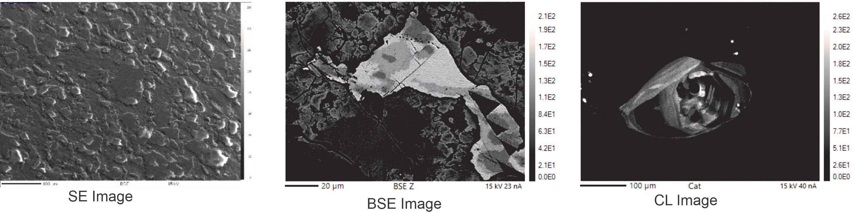
BSE Imaging: Backscattered electrons are high energy primary electrons scattered in such a direction (>90°) that they leave the target entirely. Most BSE have energies slightly lower than that of the primary electron beam. By using these emissions as video BSE images are produced and used for phase identification and mineral identification.
Cathodoluminescence (CL) Imaging: Cathodoluminescence is non-metal valence shell phenomenon leading to light emission. By using these emission as a video signal we can produce CL images that can be used to reveal the defects and impurities in materials.
X-Ray Mapping: With the help of X-Ray Mapping one can see the distribution the particular element in desired area of interest and segregation of impurities in metal alloy samples.
U-Th-Total Pb Chemical dating: Calibration Set up: 20 kV–20 nA, Quantification Set Up: 20 kV–200 nA. Following X-ray lines are used for major, trace and REE concentration as well as age determination for Monazite, viz., P–Ka, Ca–Ka, Si–Ka, Fe–Ka, Al–Ka, Y–La, La–La, Ce–La, Pr–Lb, Nd–Lb, Sm–Lb, Gd–Lb, Er–La, Eu–La, Dy–Lb, Ho–Lb, Pb–Ma, Th–Ma and U–Mb. Detailed analytical protocol is given in “Chemical dating of monazite: Testing of analytical protocol for U–Th–total Pb using CAMECA SXFive tactis EPMA at the National Centre for Earth Science Studies, Thiruvananthapuram, India,” by Sorcar et al., J. Earth Syst. Sci. (2021)130:234 (https://doi.org/10.1007/s12040-021-01738-4)
Contact:
Dr. Nilanjana Sorcar
Scientist-In-Charge
T: 0471-2511612
E: This email address is being protected from spambots. You need JavaScript enabled to view it.