

 RTI Act
- You are here:
-
Home

-
Discussion Forum
- Uncategorised
Uncategorised
Dr. Prajith A
Scientist B, Marine Geoscience Group (MGG)
Email : prajith[dot]a[at]ncess[dot]gov[dot]in
Phone(Off) : 0471-2511702
Fax : 0471-2442280
Education:
| Ph.D | : | 2016. Marine Science, CSIR-National Institute of Oceanography, Goa/Goa University |
| M.Sc | : | 2009, Geology, Kannur University, Kerala |
| B.Sc | : | 2007, Geology, Kannur University, Kerala |
Professional career:
- Scientist B, NCESS (2021 - Present)
- Project Scientist, National Centre for Polar and Ocean Research, Goa, India (2016 – 2021)
- JRF and SRF, CSIR-National Institute of Oceanography, Goa, India (2011 – 2016)
- Project Fellow, Department of Marine Geology, Mangalore University, Karnataka, India (2010 – 2011)
Areas of Specialization:
Sediment Geochemistry
Area of Interests:
Sedimentology, Paleoceanography, Paleoclimate, Marine mineral exploration, Diagenesis, Geochemistry, Isotope Geology, Environmental Magnetism
Memberships:
-
Life Member, Ocean Society of India
Honors/Awards/Fellowships:
-
CSIR Junior Research Fellowship in Earth, Atmospheric, Ocean and Planetary Sciences, Dec 2010.
-
CSIR National Eligibility Test (NET) lectureship in Earth, Atmospheric, Ocean and Planetary Sciences, Dec 2008.
Ongoing Projects:
Origin, evolution and paleoclimatic implications of estuarine-continental margin sediments (MoES-Ministry of Earth Sciences, Govt. of India)
Paleo-vegetation changes and burial efficiency of organic carbon in different depositional conditions in the Bay of Bengal and Andaman Sea during Late Quaternary and their environmental implications. DST-SERB Project.
Scientific Cruise Expeditions:
-
Participant, Dredging and Sediment core sampling in the Bay of Bengal and Andaman Sea (R.V. Sagar Kanya 373, 21.09.2021 – 15.10.2021.
-
Chief Scientist, Geological sampling, and Mapping programme in the Andaman Region (R.V MGS Sagar 30, 30.09.2019 – 07.11.2019).
-
Deputy chief scientist, Geoscientific studies in Indian EEZ area (R.V. Sagar Kanya 356, 22.02.2019 - 14.03.2019).
-
Participant, Hydrothermal Exploration in CIR/SWIR Region (R.V. MGS Sagar 24, 19.02.2018 – 02.04.2018).
-
Chief Scientist, Geological sampling, and Mapping programme in Bay of Bengal (R.V. MGS Sagar 20, 22.09.2017 – 23.10.2017).
-
Chief Scientist, Geological sampling, and Mapping programme in Bay of Bengal (R.V. MGS Sagar 16, 06.05.2017 – 12.06.2017).
-
Participant, Hydrothermal Exploration in CIR/SWIR Region (R.V. MGS Sagar 13, 12.01.2017 – 09.02.2017).
-
Participant, Geological sampling, and Mapping programme in the east coast of India (MGS Sagar 11, 12.11.2016 - 15.12.2016).
-
Participant, Geological sampling, and Mapping programme in Arabian Sea (R.V. MGS Sagar 08, 30.07.2016 – 23.08.2016).
-
Participant, Multibeam data acquisition on the western coast of India (R.V Sindhu Sankalp- 82, 12.11.2015 – 26.11.2015).
-
Seawater and sediments sampling in the Bay of Bengal (R.V. Sindhu Sadhana – 02, 23. 08.2014 – 03.09.2014).
Isotope Geochemistry Facility
NCESS has established an advanced analytical facility for isotope geochemistry and geochronology studies. The facility incorporates ICP-MS and MC-ICPMS which can be integrated with a Laser Ablation Microprobe (LAM). This combination of instruments can provide major and trace element abundances, isotopic characterisation, in-situ U-Pb dating and measurement of precise hafnium isotope ratios of various accessory mineral phases in rocks.
Instrumentation
Teledyne CETAC LSX-213 G2+ laser ablation system: The Teledyne CETAC LSX-213 G2+ laser ablation system offering a 5ns shot pulse is capable of generating > 3 mJ/pulse of laser energy with a spot size range between 4-200 μm. This allows improved absorption of energy and less transmission of the beam into the underlying epoxy. This aids in retarding the instability and U-Pb fractionation during spot analysis which indeed reduces the stabilization period allowing a short warm-up time. The laser is characterized by a flat-topped beam energy profile with pulse width < 5 nsec, enabling a repetition rate up to 20 Hz. The system is equipped with a HeLEx II sample ablation cell allowing automated stage translation with a precision of ±2–3 μm for pre-setting of analytical points. The HeLEx II with an internal volume cup enables effective sample transportation to the ICPMS through the articulated gas inlet/outlet arm. The ablated material from the sample is pushed out of the chamber by purging helium gas which later mixes with the carrier argon gas and gets transported into the plasma. The fast washout of the laser ablation system allows rapid measurement of elements with a minimal fractionation effect.
Agilent 7800 Quadruple ICPMS: The Agilent 7800 Quadrupole ICPMS is equipped with a dual-mode discrete dynode electron multiplier as an ion detector. The quadrupole mass analyzer is a 3 MHz hyperbolic rod having a mass range 2–260 u and variable mass resolution from 0.3 u to 1.0 u. This enables a sensitivity ≤ 5 × 10⁻⁷ counts for low masses and ≤ 1 × 10⁻⁷ counts for higher masses. The Scott-type double-pass quartz spray chamber with High Matrix Introduction system tandem with 500 - 1600W RF generating capability allows complete ionization of the introduced sample. The Octopole Reaction System (ORS) with He gas in the cell interacts with the ions from the sampling cone to avoid the interference of polyatomic molecules.
Nu Plasma III Multi collector ICPMS: Nu-PLASMA-III is a double-focusing Multi Collector ICP Mass Spectrometer (MC-ICP-MS) designed to provide a high precision isotope ratio of elements. The MC-ICPMS is equipped with 16 Faraday detectors, 2 Daly detectors and 4 Ion counters. The instrument retains the unique, patented, variable dispersion Zoom lens enabling the simultaneous measurement of the isotopes of elements from lithium to the actinide series on its static collector arrays. This allows comprehensive coverage of different isotopic applications over the full mass range.
Calibration strategy
U-Pb geochronology using LA-ICPMS: The ICPMS is tuned for U-Pb geochronology by ablating a raster over NIST 610 (Pearce et al., 1997) standard glass reference material to get the maximum signal intensity and stability for analyzed masses. The ICPMS parameters along with nebulizer and laser gas flows are adjusted to achieve maximum counts for the monitored masses keeping U/Th ratio ~1. Plasma oxidation level is monitored by Th/ThO ratio and double charged ion ratio was monitored using Ca/Ca2+. The He flow is optimized to get maximum counts and to minimize fractionation during sample transport. Spot analysis on accessories is conducted at a spot size of 30 microns with a laser fluence of ~4 J/cm2 on the sampling surface and a repetition rate of 10 Hz in Q-switch mode. LIEF correction is applied using zero ablation time intercepts of least-squares linear regression lines fitted to the time-resolved isotopic ratio data using the protocol of Košler and Sylvester (2003) whereas inter-element fraction and instrumental mass discrimination are corrected by normalization to a matrix-matched primary reference material, which is analyzed during the analytical sessions under the same conditions as the samples. In-situ trace element determination is carried out using NIST 612 and NIST 610 (Pearce et al., 1997) as reference standards for time-drift correction and quality monitoring. Off-line data reduction is carried out using Iolite 4.1, ICPMS DataCalc 12.0 and Glitter software.
Lu-Hf measurements in zircons using LA-MC-ICPMS: Zircons analyzed for U-Pb dating will be reanalyzed and the laser spots positioned as close as possible to the previous crater. Before integrating with the LA, pure 25 ppb JMC 475 solution is analyzed for calibration and peak selection. For hafnium measurement, the laser is operated at a spot size of 50 μm at 10 Hz repetition rate in Q-switch mode, imparting a laser fluence of ~3 J/cm2 in the sample surface. Data are reduced off-line and calculated isotopic ratios and elemental concentrations are processed using Iolite 4.1. Isotopic fractionation of Hf is determined by measuring interference-free 179Hf/177Hf and βHf is determined using a constant 179Hf/177Hf value of 0.7325 (Patchett et al.,1981). Similarly, isotopic fractionation of Yb is corrected by calculating βYb using the measurement of interference-free 173Yb/171Yb using a constant 173Yb/171Yb value of 1.132685 (Vervoort et al., 2004). Correction for the interference of 176Yb on 176Hf is carried out by measuring the interference-free 173Yb and using the constant 176Yb/173Yb value of 0.796218 (Chu et al., 2002).
Detailed analytical protocol for all these techniques is according to “Dev, J. A., Tomson, J. K., Sorcar, N., & Nandakumar, V. (2021). Combined U-Pb/Hf isotopic studies and phase equilibrium modelling of HT-UHT metapelites from Kambam Ultrahigh temperature Belt, South India: Constraints on tectonothermal history of the terrain. Lithos, 106531 (DOI; https://doi.org/10.1016/j.lithos.2021.106531) .”
Sample Preparation
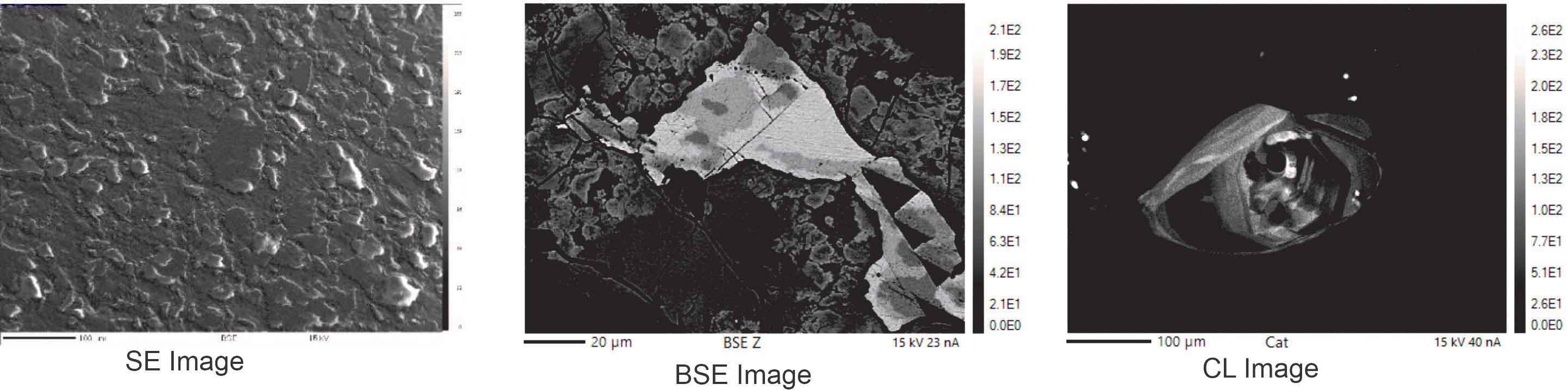
Accessory minerals are analyzed in both mineral separates as well as in thin sections. Accessory minerals are separated from bulk rock samples using conventional Wilfley table and magnetic separation methods in the Thin section and Sample preparation Laboratory at NCESS. The handpicked grains are mounted in epoxy-filled blocks of 2.5 cm diameter and subsequently sectioned and polished. Grain mounts are later cleaned in different steps with ethanol and deionized water in an ultrasonic bath to remove surface contamination during sample preparation before insertion into the laser sample cell. This allows skipping pre-ablation during LA analysis even though pre-ablation is carried out in rare cases if surface contamination is noticed. The internal structure of targeted minerals is studied using backscattered electron (BSE) and cathodoluminescence (CL) images taken either from SEM or EPMA laboratories at NCESS.
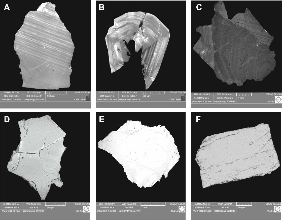 Figure: SEM images of different natural reference standards. A. 91500 Zircon, B. Plesovice Zircon, C. BB11 Zircon, D. R632 Rutile, E. Diamantina Monazite, F. IN1 Baddeylite
Contact:
Dr. Tomson J Kallukalam
Scientist-In-Charge
T: 0471-2511655
E: This email address is being protected from spambots. You need JavaScript enabled to view it.
Electron Probe Micro Analyser (EPMA) Facility
NCESS has installed a Fifth Generation EPMA (Electron Probe Micro Analyser) SXFive-Tactis from CAMECA, France. EPMA is an instrument primarily used for the in situ non-destructive chemical analysis of minute solid samples. The ability of EPMA to acquire precise, quantitative elemental analyses at very small "spot" sizes (1-2 microns), mainly by wavelength-dispersive spectroscopy (WDS) combined with the ability to create detailed images of the sample, makes it possible to analyze geological materials in situ and to resolve complex chemical variation within single phases. NCESS-EPMA will be primarily used for (i) chemical analysis of geological materials, (ii) analysis of individual phases (e.g., igneous and metamorphic minerals), (iii) U-Th-Pb geochronology of minerals such as monazite, zircon etc.
Instrumentation
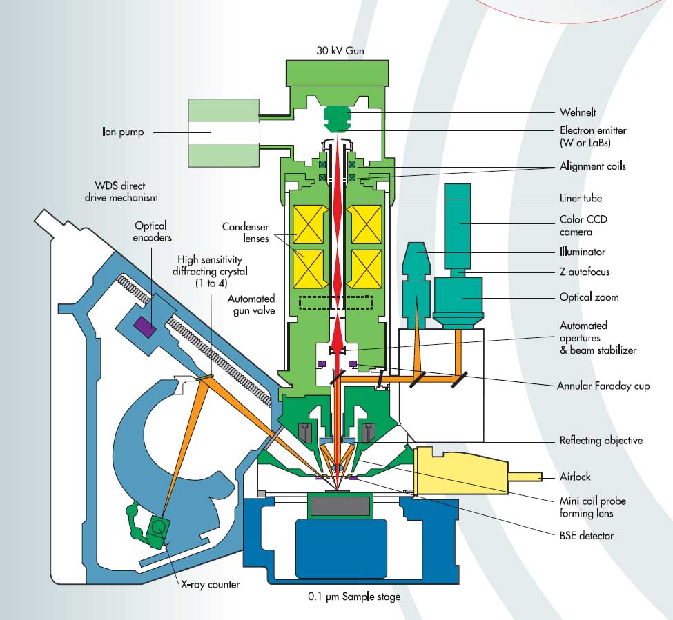
NCESS-CAMECA SXFive-Tactis is equipped with five wave length dispersive spectrometers, BSE detectors, SE detectors, cathodoluminescence and sophisticated visible light optics. The self-biased electron gun of the instrument has the provision for both W and LaB6 filaments. The instrument is configured in such a way so that operation as well as basic imaging and data processing are made easy for having intuitive touch screen interface. In expert mode, the interface is designed to have benefit from a full complement of different tool parameters and software options. The system involves a high degree of automation with the capacity of prolonged unattended analytical schedules with high analytical precision, accuracy and reproducibility with minimum down-time, high quality imaging at high spatial resolution including multi-element x-ray mapping. Vertical WDSs are compatible to have suitable combinations in high sensitivity diffracting crystals (e.g. LTAP, LPC1, LLIF and LPET) as well as six diffracting crystals such as TAP, LIF, PET, PC0, PC3 and PC2. Another advantage of this instrument is that it is equipped with a fully integrated Electron Dispersive Spectrometer hypermapping module making data processing and analysis faster and easier. SX- Five with LaB6 Column: Accelerating Voltage: 15KV and 20 KV; Beam Current: 20 uA onwards; Beam Stability: +-0.5% per hour @ 20kV, 20nA.
Capabilities
Qualitative analysis: WDS Spectrum-to find out the elements present. Wavelength Dispersive Spectrometry is acknowledged as the method of choice for high precision quantitative microanalysis. The SXFive-Tactis spectrometers have the highest resolution and, as a result high peak to background ratios, thereby providing high sensitivities. The absolute spectral positioning is provided by optical encoders attached to the system. Only one peak measurement is needed to calibrate the entire spectrometer range.
Quantitative analysis: Accurate quantification of chemical composition of any solid material. SXFive-Tactis delivers the true quantitative analysis. The quantitative data can be displayed such as wt%, oxide wt%, cations. See the table below for our precision:
|
Cameca Silicate Std (Alamandine) |
sample |
Cameca Sulphide Std (Pyrite) |
sample |
Uranium Oxide Std |
sample |
|||
|
SiO2 |
37.87 |
37.51 |
S |
53.06 |
52.86 |
Al2O3 |
0.29 |
0.07 |
|
TiO2 |
0.07 |
0.04 |
Fe |
46.51 |
45.89 |
UO2 |
89 |
89.28 |
|
Al2O3 |
21.36 |
21.16 |
Co |
0.5 |
0.35 |
SiO2 |
2.3 |
7.23 |
|
Cr2O3 |
0.0 |
0.03 |
Ni |
0.01 |
0.01 |
ZrO2 |
1.09 |
0.3 |
|
FeO |
34.6 |
30.77 |
Cu |
0.02 |
0.06 |
TiO2 |
1.11 |
0 |
|
MnO |
0.37 |
1.64 |
Zn |
0.01 |
0 |
Gd2O3 |
0.27 |
0.89 |
|
MgO |
2.34 |
9.41 |
As |
0.02 |
0.04 |
PbO |
2.0 |
1 |
|
CaO |
4.34 |
0.65 |
- |
- |
CaO |
1.88 |
0.04 |
|
|
Na2O |
0.10 |
0.0 |
- |
- |
Y2O3 |
0.61 |
0.24 |
|
|
K2O |
0.0 |
0.0 |
- |
- |
La2O3 |
0.18 |
0.14 |
|
|
ZnO |
0.12 |
0.12 |
- |
- |
Ce2O3 |
1.21 |
0.63 |
|
|
P2O5 |
0.06 |
0.01 |
- |
- |
Nd2O3 |
0.86 |
0.49 |
|
|
Total |
101.23 |
101.34 |
99.669 |
99.21 |
Total |
100.8 |
100.31 |
BSE Imaging: Backscattered electrons are high energy primary electrons scattered in such a direction (>90°) that they leave the target entirely. Most BSE have energies slightly lower than that of the primary electron beam. By using these emissions as video BSE images are produced and used for phase identification and mineral identification.
Cathodoluminescence (CL) Imaging: Cathodoluminescence is non-metal valence shell phenomenon leading to light emission. By using these emission as a video signal we can produce CL images that can be used to reveal the defects and impurities in materials.
X-Ray Mapping: With the help of X-Ray Mapping one can see the distribution the particular element in desired area of interest and segregation of impurities in metal alloy samples.
U-Th-Total Pb Chemical dating: Calibration Set up: 20 kV–20 nA, Quantification Set Up: 20 kV–200 nA. Following X-ray lines are used for major, trace and REE concentration as well as age determination for Monazite, viz., P–Ka, Ca–Ka, Si–Ka, Fe–Ka, Al–Ka, Y–La, La–La, Ce–La, Pr–Lb, Nd–Lb, Sm–Lb, Gd–Lb, Er–La, Eu–La, Dy–Lb, Ho–Lb, Pb–Ma, Th–Ma and U–Mb. Detailed analytical protocol is given in “Chemical dating of monazite: Testing of analytical protocol for U–Th–total Pb using CAMECA SXFive tactis EPMA at the National Centre for Earth Science Studies, Thiruvananthapuram, India,” by Sorcar et al., J. Earth Syst. Sci. (2021)130:234 (https://doi.org/10.1007/s12040-021-01738-4)
Contact:
Dr. Nilanjana Sorcar
Scientist-In-Charge
T: 0471-2511612
E: This email address is being protected from spambots. You need JavaScript enabled to view it.
Awards
| Sl.No | Name of Award | Awardee Details | ||
| 1 | Certificate Of Merits for Scientists/Engineers | 
|
Dr. S. Kaliraj, Scientist C, Crustal Dynamics Group. | |
Main Chemical Lab (MCL)
Main Chemical lab is equipped to carryout routine evaluation of chemical and physical parameters in water and wastewaters for both qualitative and quantitative determination of light metals and pesticides content.Consisting of LC-MS/MS, GC-MS/MS, MP-AES, GC, UHPLC, AAS, Continuous Flow Analyser (CFA), UV-Vis-NIR Spectrophotometer, Flame photometer, CHNS Analyser, TOC Analyser, Surface Area Analyser, Sedigraph, Mercury Analyser, IC.
Instruments available are:
- GC – Perkin Elmer (N651-9101 CLARUS 500)
- Bacteriological Incubator – Labline
- Laboratory oven – Labline
- Muffle Furnace – Bio-Technics India
- BOD Incubator – Calton
- UV Spectrophotometer – Shimadzu (UV-1800)
- Flame photometer – (CL-361)
- Flame photometer – (CL-378)
- Electronic weighing balance – Sartorius (BP221 S)
- Sediment Squeezer
- Rotary Vacuum Evaporator – Eyela (NVC 2100)
Download Water/Sediment Analysis Request Form
News and Events
Research Updates
- U-Pb geochronology of rutiles from Southern granulite Terrane, India
- Metamorphic and tectonic evolution of the Southern Granulite Terrane, India
- Origin of TTGs and high-K granitoids in the Bundelkhand Craton, Central India
- Detrital zircons in crustal evolution: A perspective from the Indian subcontinent
- Age of Kaladgi Supergroup, India
- Geochemical provenance of an Indo- Arabian stone anchor from Manikapatna
- Paleo fluids in the petroliferous basins of western offshore, India
